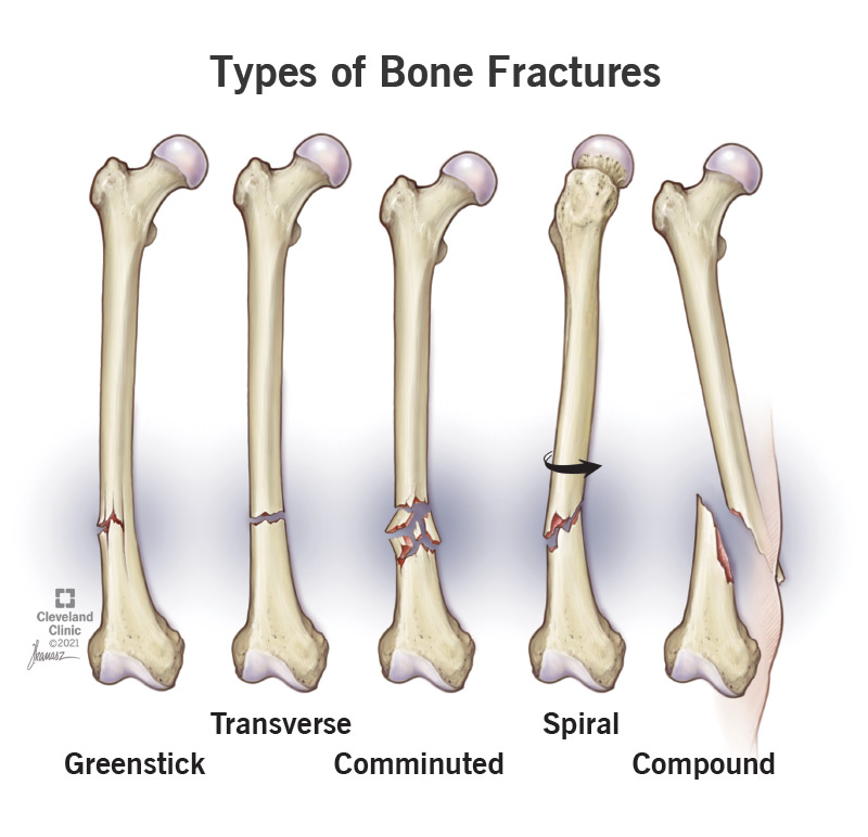
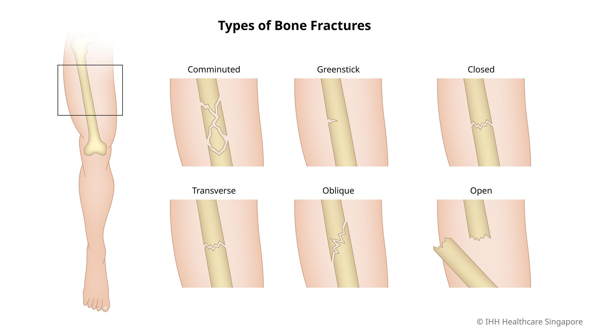
In this comprehensive article, we shed light on the intricacies surrounding comminuted fractures, exploring their causes, symptoms, and treatment options. A comminuted fracture occurs when a bone breaks into several fragments, often requiring quick and precise medical intervention. By understanding the underlying causes and recognizing the telltale symptoms, individuals can seek timely medical attention, thus maximizing their chances of successful recovery. Additionally, we delve into the various treatment approaches available, encompassing both surgical and non-surgical options, to provide a comprehensive understanding of comminuted fractures and their management.

Definition of Comminuted Fracture
What is a comminuted fracture?
A comminuted fracture is a type of bone fracture characterized by the bone being broken or shattered into multiple fragments. Unlike a simple or clean fracture where the bone breaks in one or two pieces, a comminuted fracture results in three or more bone fragments. This makes comminuted fractures more complex and challenging to treat.
How does a comminuted fracture occur?
Comminuted fractures typically occur due to high-energy trauma or severe force being applied to the bone. Common causes include car accidents, falls from a significant height, sports injuries, and crushing injuries. The greater the force exerted on the bone, the more likely it is to shatter into multiple fragments. In some cases, underlying diseases such as osteoporosis or pathological conditions can weaken the bone, increasing the risk of a comminuted fracture even with less force.
Causes of Comminuted Fractures
Trauma or accidents
Traumatic events like car accidents, sports injuries, or falls are frequent causes of comminuted fractures. The impact or force applied to the bone exceeds its strength, leading to the bone breaking into multiple fragments. The severity of the fracture will depend on the magnitude of the force and the location of the impact.
Osteoporosis
Osteoporosis is a condition that weakens the bones, making them more susceptible to fractures. When osteoporosis is present, even a minor injury or fall can lead to a comminuted fracture. This is because the bones have lost their density and become brittle, unable to withstand the force applied to them. Older adults, especially women after menopause, are at a higher risk of developing osteoporosis and experiencing comminuted fractures.
Pathological conditions
Certain pathological conditions can weaken the bones and increase the likelihood of comminuted fractures. These conditions include bone tumors, bone infections, and bone cysts. The presence of these conditions renders the bone more susceptible to fractures, particularly when any external force is applied.

Symptoms of Comminuted Fractures
Visible deformity
One of the most apparent signs of a comminuted fracture is a visible deformity at the site of the fracture. The bone fragments may be displaced or misaligned, causing an abnormal shape or bulge in the affected area. This deformity is often noticeable and can be observed even without a medical examination.
Intense pain
Comminuted fractures are typically associated with severe pain. The pain may be sharp, throbbing, or constant, depending on the individual and the location of the fracture. The intensity of the pain can make it difficult to move or use the affected limb.
Swelling and bruising
Immediately after a comminuted fracture, swelling and bruising are common. The injury triggers an inflammatory response, causing the area to swell and become tender. The presence of swelling and bruising helps to localize the fracture and may extend beyond the immediate region of the fracture.
Inability to bear weight or use the affected limb
Because of the severe pain and displacement of bone fragments, individuals with comminuted fractures often experience difficulty bearing weight or using the affected limb. This limitation in mobility is a result of the instability caused by the fracture and the discomfort it produces.
Bone fragments protruding through the skin
In some cases of comminuted fractures, the bone fragments may pierce through the skin and become exposed. This is known as an open or compound fracture and is considered a medical emergency. The protruding bone fragments increase the risk of infection and necessitate immediate medical attention.
Diagnostic Procedures for Comminuted Fractures
Physical examination
A physical examination is the initial step in diagnosing a comminuted fracture. The medical professional will assess the affected area, looking for signs of deformity, swelling, or bruising. They will also evaluate the range of motion, checking for any limitations or increased pain with movement.
X-rays
X-rays are commonly used to confirm the presence of a comminuted fracture. X-ray images allow healthcare providers to visualize the bones and identify any fractures or bone fragments. This imaging technique helps determine the exact location of the fracture and guides treatment decisions.
Computed Tomography (CT) scan
A CT scan provides detailed cross-sectional images of the bones, allowing for a more comprehensive assessment of the fracture. It can help identify the extent of the fracture, the size and displacement of the bone fragments, and any associated injuries. CT scans are particularly helpful in complex comminuted fractures where a clear understanding of the fracture pattern is crucial for treatment planning.
Magnetic Resonance Imaging (MRI)
MRI scans may be ordered if there is a suspicion of soft tissue or ligament damage in addition to the comminuted fracture. MRI provides detailed images of the soft tissues, such as muscles, tendons, and ligaments, allowing for a more comprehensive evaluation of the entire injury.
Bone scan
A bone scan may be recommended in cases where a comminuted fracture is suspected but not clearly visible on other imaging studies. This diagnostic test involves injecting a small amount of radioactive material into the bloodstream, which binds to areas of increased bone activity. A special camera then detects this radiation and produces images that can help identify fractures or bone abnormalities.

Classification of Comminuted Fractures
Type I: Simple wedge fracture
Type I comminuted fractures are relatively stable fractures with one large wedge-shaped fragment and smaller, non-displaced fragments. These fractures typically occur in long bones, such as the femur or tibia, and are often amenable to non-surgical treatment methods.
Type II: Butterfly or H-type fracture
Type II comminuted fractures, also known as butterfly fractures or H-type fractures, involve a central butterfly-shaped fragment with separate displaced fragments on either side. This fracture pattern can cause significant instability and may require surgical intervention for proper alignment and fixation.
Type III: Multi-fragmentary fracture
Type III comminuted fractures involve three or more fragments, resulting in significant bone displacement and instability. These fractures are the most severe and require surgical intervention to restore proper alignment and stability.
Treatment Options for Comminuted Fractures
Non-surgical treatment
Non-surgical treatment may be considered for stable comminuted fractures that maintain proper alignment. This typically involves immobilization with casts or braces to restrict movement and support the healing process. Pain management is also an essential component of non-surgical treatment, often involving medications and physical therapy to regain strength and mobility.
Surgical treatment
Surgical treatment is often required for comminuted fractures to realign the bone fragments and promote proper healing and stability. Different surgical techniques may be used depending on the specific fracture pattern and location. The primary goals of surgery are to reconstruct the bone, restore stability, and minimize the risk of complications.
Internal fixation
Internal fixation involves the use of various implants, such as screws, plates, rods, or intramedullary nails, to stabilize the fractured bone and facilitate healing. These devices are surgically inserted and provide support and alignment during the healing process. Internal fixation is commonly used in comminuted fractures where the bone fragments can be adequately aligned and secured.
External fixation
External fixation utilizes a stabilizing device positioned outside the body to stabilize and immobilize the fractured bone. The device consists of pins or wires that are inserted into the bone and connected to an external frame. This technique is often used in more complex comminuted fractures where internal fixation may not be feasible or sufficient to achieve stability.
Joint replacement
In cases where the comminuted fracture involves a joint and severely affects its function, joint replacement surgery may be necessary. Joint replacement involves removing the damaged joint surfaces and replacing them with artificial joint components. This procedure aims to restore joint function while addressing the fracture and any associated damage.
Recovery and Rehabilitation Process
Immobilization with casts or braces
Following a comminuted fracture, immobilization with casts or braces is essential to promote healing and stability. These devices restrict movement and protect the affected area from further injury. The duration of immobilization will vary depending on the severity and location of the fracture, with regular follow-up appointments to monitor progress and ensure proper healing.
Physical therapy
Physical therapy plays a critical role in the recovery and rehabilitation process for individuals with comminuted fractures. A customized rehabilitation program is designed to help restore range of motion, strength, and function. Physical therapists guide patients through exercises, stretches, and functional activities to promote healing, prevent muscle atrophy, and improve overall mobility.
Pain management
Pain management is an integral part of the recovery process for comminuted fractures. Various pain management techniques may be employed, including medications, cold or heat therapy, immobilization, and physical therapy. Pain management not only enhances patient comfort but also facilitates participation in rehabilitation activities.
Resumption of daily activities
The resumption of daily activities will depend on the severity and location of the comminuted fracture, as well as the individual’s healing progress. Gradual reintroduction of activities, such as walking, lifting, or sports, is typically guided by healthcare professionals to ensure proper healing and minimize the risk of reinjury.
Complications Associated with Comminuted Fractures
Delayed healing
Comminuted fractures pose a higher risk of delayed healing compared to other types of fractures. The complexity of the fracture and the presence of multiple bone fragments can impede the healing process, resulting in a lengthened recovery timeline. Factors such as age, overall health, and the presence of underlying medical conditions can also influence the healing process.
Malunion or nonunion
Malunion occurs when the fractured bones heal in a misaligned or abnormal position. Nonunion refers to the failure of the bone fragments to heal together. Both complications can result from the inability to achieve proper alignment or stability during treatment. Surgical intervention may be necessary to correct malunion or nonunion and restore proper bone healing.
Infection
Infection is a potential complication associated with comminuted fractures, particularly for open fractures or those requiring surgical intervention. The exposure of bone fragments to the external environment increases the risk of bacterial contamination. Prompt treatment with antibiotics and appropriate wound care is crucial to prevent or manage infections.
Nerve or blood vessel damage
Comminuted fractures can potentially damage nearby nerves and blood vessels, leading to sensory or motor deficits. The bone fragments can exert pressure or cause direct injury to these structures, resulting in decreased sensation, muscle weakness, or compromised blood flow. Early recognition and appropriate treatment are necessary to minimize the long-term consequences.
Prevention and Tips for Avoiding Comminuted Fractures
Fall prevention measures
Many comminuted fractures occur as a result of falls. Taking preventive measures to reduce the risk of falls can help avoid such fractures. These measures include maintaining a safe and obstacle-free environment, using handrails on stairs, wearing appropriate footwear, and addressing any hazards that may contribute to falls.
Maintaining bone health
Strong and healthy bones are more resistant to fractures, including comminuted fractures. Taking steps to maintain bone health can help reduce the risk of fractures. This includes eating a calcium-rich diet, getting regular exercise, quitting smoking, limiting alcohol consumption, and ensuring an adequate intake of vitamin D.
Wearing protective gear
For individuals participating in activities with a higher risk of trauma, wearing protective gear can provide an extra layer of protection. Helmets, knee pads, elbow pads, and other sports-related protective gear can help prevent and reduce the severity of injuries, including comminuted fractures.
Prognosis for Comminuted Fractures
Factors affecting prognosis
Several factors can influence the prognosis of comminuted fractures. The location, severity, and type of fracture, the individual’s age and overall health, the presence of underlying medical conditions, and the treatment received all play a role in determining the outcome. Prompt and appropriate treatment, adherence to rehabilitation programs, and close monitoring by healthcare professionals are essential for a favorable prognosis.
Expected recovery timeline
The recovery timeline for comminuted fractures varies depending on individual factors and the specific details of the fracture. Generally, healing can take several months, and rehabilitation may continue for an extended period. Regular follow-up appointments with healthcare providers and adherence to treatment and rehabilitation protocols are crucial for achieving the best possible recovery outcome.


